More aboutPtosis...
More About Ptosis - Why, Who, When
Why does ptosis ( droopy eyelids) occur?
Droopy eyelids can be caused by the following-
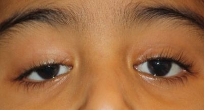
a. Congenital - Since birth. The droopy eyelid can block the pupil of the eye, obstruct the vision, and hamper the development of vision - a condition called lazy eye or amblyopia. The initial treatment is by giving glasses and patching the good eye to develop the strength of the ptotic droopy eye. The next step is surgery of the ptosis.
b. Adult ptosis can be due to weakening of the muscles of the eyelid. This may be due to increasing age, an injury, many years of chronic eye rubbing such as in allergic conjunctivitis, or many years of use of contact lens.
c. Adult onset ptosis can be due to onset of a disease process of the eyelid muscles or eye nerves. Some examples are CPEO, 3rd nerve paralysis, or myasthenia.
Ptosis Surgery - Best Choice
Surgery for ptosis ( droopy eyelids) can be done by different techniques.
Step1. We rule out the diseases of the eye mentioned in the previous section. These need special consideration. This is by clinical examination, as well as blood test etc.
Step 2. We measure the strength of the eye eye muscle, the amount of ptosis, and the movement capability of the eye.
Step 3.
a.If the strength of the eye muscle is good, we do a levator surgery- we tighten the muscle inside the eyelid to make it work more effectively. This can also be variously called levator plication, levator reattachment or levator resection. This is done through an incision hidden in the eyelid crease.
b. If the muscle strength is good, with a small amount of ptosis , we can do Muller's Muscle Conjunctival resection ( MMCR). This is done from inside the eyelid.
c. If the eyelid muscle is weak, we attach it to the forehead muscle from the inside, so that the eye can be opened using the strength of the forehead muscle. There will be 3 tiny puncture marks near the eyebrow.The attachment can be using a synthetic material like silicone sling, or a natural graft called fascia lata. A silicone sling surgery is quicker, can be done under local anesthesia, but there is the placement of an artificial material within the eyelid. The fascia lata sling surgery takes longer, needs general anesthesia, there is an additional cut on the leg of the patient. However, fascia lata sling is considered the gold standard in sling surgery, with long term proven results.
Your surgeon will determine the technique of the surgery which is most appropriate for you.
Limitations of ptosis surgery
1. Results of ptosis surgery are not fully predictable. While the surgery is performed after taking the measurements, and using the appropriate technique ( see above), each person's body is unique, and heals in a different way. In case the operated eyelid is 2 mm or more different from the other, a repeat surgery may be needed. About 2 in 10 patient require repeat surgery.
2. One should expect an incomplete eye closure after ptosis surgery. It is initially treated with tear substitutes. In most patients, their eyes tolerate this incomplete closure well. Very rarely, the eye does not tolerate the gap in closure, and can develop wounds on the cornea. This will need treatment.
3. The target of ptosis surgery is to make the eyelids similar when looking straight ahead. There will always be a difference int he appearance of the two eyes when the patient looks up or looks down.
The limitations are not complications of ptosis surgery, it is the nature of the surgery.
More about Watering Eye
What is tear duct block in a child? What is the treatment for watering of the eye in a child?
Tear duct block in a child is also called congenital naso-lacrimal duct obstruction ( CNLDO). The child starts to have watering, stickiness and discharge from the eye, usually a few weeks after birth. The condition can affect one eye or both eyes.
The treatment is done is steps.
Antibiotic drops can improve the current infection, but will not help is actual cure of the block
If your child is less than a year old, your doctor can advise massage near the corner of the eye. Please make sure you understand the CORRECT technique of the massage. A large number of children will recover with regular lacrimal sac massage.
If your child is more than a year old, has excessive dirt and discharge from the eye, or is scheduled for other eye surgeries, your doctor will advise a small surgery called probing. Most babies will recover after probing, but in a few, the tear duct can close down again.
In case the tear duct in a child closes again after probing, additional placement of a silicone tube ( lacrimal intubation) or an open surgery will be required.
How is tear duct block diagnosed?
l Lacrimal sac syringing is the commonest test for tear duct block. Your doctor uses a fine tube to push some water into your tear duct in the corner of the eye. If you feel the water in the nose or the throat, your tear duct is open. The open duct gives a’Patent’ syringing. Syringing is a test, it is NOT a treatment. If you do not feel the water in the nose or throat, the tear drainage duct is blocked.
l If there is a swelling inthe inner corner of the eye, your doctor can press over it. If pressure over it causes dirt and discharge to come out, the tear duct block is confirmed. This is called regurgitation on pressure over the lacrimal sac.
l Your doctor may placea tiny amount of orange coloured dye called fluorescein in your eye. This is a very safe procedure. After waiting 5 minutes, your doctor will check the eye. If the dye has drained away, your tear duct is open. If the dye is still present in the eye, the tear duct is blocked. This is the Fluorescein Dye Disappearance Test (FDDT).
l In rare, complex conditions, a CT scan with radio opaque dye may be required. This may needed in patients with history of previous injury in the area. This is called the CT DCG.
What is dacryocystorhinostomy?
Dacryocystorhinostomy (DCR) is a surgery to correct a blocked tear drained duct . The doctor makes a new passage for the tears to drain into your nose.
Why do I have watering eye even when the syringing is patent?
Watering of the eye can be due to many reasons.
Block of the tear drainage duct is one of the common causes of the watering of the eye.
Allergic eye disease, meibomitis or eye infections can cause watering of the eye, These are treated with medicines.
Some eyelid conditions such as entropion, eyelashes rubbing on the eye surface, paralysis of the eyelid, difficulty closing the eye, eyelid turned outwards ( ectropion) - call all cause watering of the eye. Most of these conditions are treated by surgery.
More About Eye Cancers

What are the types of eye cancer? What happens in eye cancer?
Cancer can occur in various parts of te eye - the eyelid, the conjunctiva ( white part of the eye), behind the eye ( tumor of the orbit), inside the eye.
There can be be a growth or a lump in the different parts of the eye, which will keep increasing. Eventually it can damage the entire eye, damage the vision, and even spread to other parts of the body . This is a primary tuor of the eye.
In some patients, who have cancer in another part of the body, the disease can spread to the eye. This is a secondary eye tumor, also called a metastasis.
What is OSSN? What is the treatment of OSSN?
Conjunctival squamous cell carcinoma is also called OSSN ( ocular surface squamous neoplasia). It is a tumor of the conjunctiva , the white part of the eye.
It starts as pink or whitish swelling on the surface of the eye , the white part, the black pat, or both. There may be some irritation. If it grows to cover the central part of the eye, the vision will be blurred.
Your doctor may start you on eye drops to treat the OSSN. The eyedrops can be interferon alpha 2B, mitomycin C or 5 fluorouracil. These are not available commercially. Your doctor will reconstitute them for you, from available injections. These have to be stored in the refrigerator. You will have to visit your doctor every 2 weeks, to check on the improvement, and also to have the drops freshly mixed.
In some cases, your doctor will decide to treat your OSSN by surgery. OSSN Surgery should follow a protocol - surgery with clear margin, cryotherapy applied to edges of surgical site, and sample to be sent for laboratory testing for histopathology.
Even after treatment, your doctor will give you a schedule for follow up, to check if the tumor is coming back.
When OSSN is treated early and by the correct method, the results are very good.
What does an eyelid cancer look like? What is the treatment of eyelid cancer?
The common eyelid cancers in India are sebaceous carcinoma, basal cell carcinoma and squamous cell carcinoma. https://www.ncbi.nlm.nih.gov/pmc/articles/PMC7774165/ Dr Roshmi Gupta has published in the Indian Journal of Ophthalmology about her experience with eyelid cancers.
Eyelid cancer starts as a small lump on the eyelid, which keeps growing. It may be pink, red or dark in colour. There may be some itching or irritiation, but often it has no discomfort.
if left untreated, it can extend behind the eye, cause loss of eye and vision, or even spread to the other parts of the body, endangering the life of the patient.
The doctor will operate on the eyelid tumor, removing it completely, with clear margins. She will send the sample for biopsy and histopathology to confirm. next, she will perform eyelid reconstruction to repair the site of eyelid surgery.
Even after treatment, your doctor will give you a schedule for follow up, to check if the tumor is coming back.